Plant Cell Wall
A rigid layer of a complex polysaccharides found, on the outer surface of the plasma membrane, in plant cells, is called cell wall, which is one of the most important characteristic feature of plants. If plants cell wall is digested by cellulase and pectinase enzymes the result is cells free of cell wall. It is called protoplast and it is always spherical is shape. Using compound microscopes it is possible to distinguish different wall layers by specific stains. They are middle lamellae, primary wall, secondary and tertiary wall.
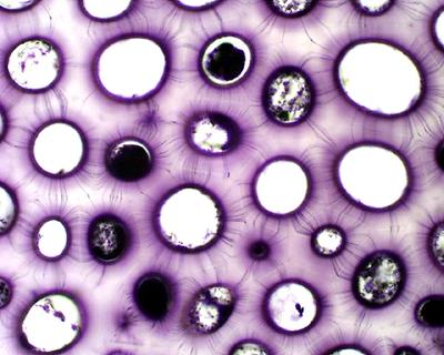
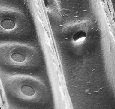
Invariably the cell wall layers are perforated all over; these areas are called pits. Basing on the structural organization of pits they are classified into simple pits and bordered pits. It is through the pits a large number fine cytoplasmic strands pass through and act as interconnecting system for the inter cellular movement of cellular components; thus provide a continuum among the cells, such strand are called protoplasmic strands or plasmodesmata, a living membrane tubular system. These strands operate as long cells are living, when they are dead they leave holes.
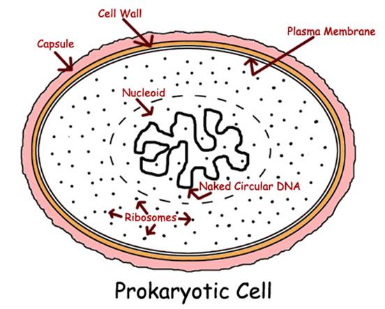
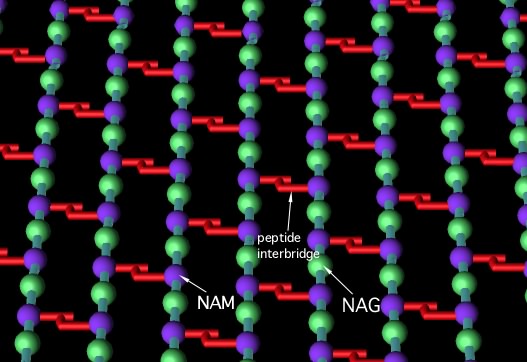
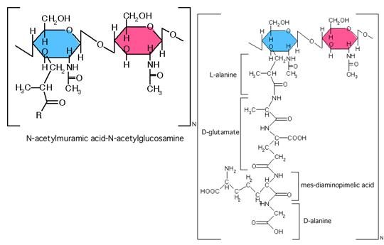
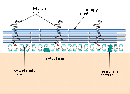
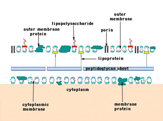
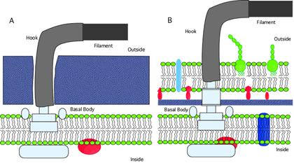
In the case of bacteria the cell walls are of two different kinds; the one is gram negative (ex. E.coli) and the other is gram positive (gram positive (ex. Bacillus subtilis). In the case of Gram negative bacteria, out side the plasma membrane, one finds periplasmic space which is filled with few layer of peptidoglycans wall. Next to it one finds another layer of lipoprotein membranous layer decorated with a variety of polyglycosylated proteins or and with lipopolysaccharides.

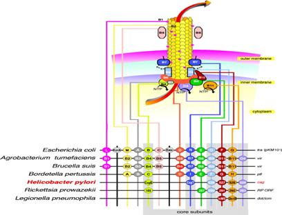
Agrobacterial componenets compared with other type of bacteria
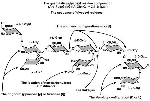
Chemical composition and the structure of primary wall:
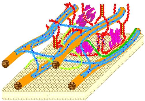
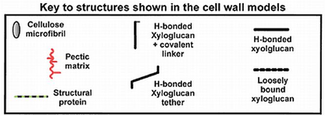
Basically the primary wall is made up of cellulose and hemicelluloses. While pure cellulose acts as crystalline structures, hemicelluloses like polyarabons, polyxylans, polymannans and polygalactans act as the paracrystalline amorphous materials. Added to these materials polypeptide chains called extensins, rich in hydroxproline amino acids are also found. Extensin acts as cross links between cellulose complex and non cellulose fibrils. Most of the primary cell walls contain more or less 90% of polysaccharides and 10% proteins. Among them, extensin is a glycoprotein with mol. wt of 40KD. It is rich in hydroxyproline and few other amino acids present are serine, Lysine, Histidine and tyrosine. Though extensin acts as a cross link between the cellulosic fibers and non cellulosic heme-cellulose, extensins, interconnect them by chemical cross links. A structural model was built on the basis of X-ray diffraction pattern and enzymatic studies.
Micro fibrils inturn organize into bigger bundles called macro fibrils. To begin with micro fibrils of 6.2 mm thickness are deposited randomly during primary wall formation and gradually the orientation of the fibrils changes to either longitudinal transverse or obliquely orienting. However, these fibers are deposited layers after layers. The interspaces are later filled with other materials like hemicelluloses, lignins, tannins, fatty acids and other related compounds. The impregnation of such materials provides fortification and rigidity to the cell wall layers.
Biogenesis of cell wall:
Biosynthetic activity of cellulose is very rapid during the cell division and early growth of cells. The enzymes responsible for the synthesis of cellulose and non-cellulosic components are found in rough endoplasmic reticulum. Then they are transported into the lumen of smooth endoplasmic, which inturn pinch of vesicles containing enzyme. Such vesicles fuse with one another at the proximal face of Golgi apparatus. Thus cell wall precursors find their way into golgi membranes where the enzymes take over the functions of modification of precursors. Then they are processed and packed into trans golgi vesicles. Later such vesicles are pinched off the transported towards plasma membranes, where vesicles fuse with membranes and release the enzymes and other raw materials for the cell wall. Microtubules and microfilaments play a significant role in the transportation and deposition of vesicles at the vicinity of plasma membranes.
It is at the outer surface of the plasma lemma, cellulose synthetases become active and start synthesizing cellulose fibrils, which soon organize into micelles. With the input of other non-cellulosic component micelle inturn associate into micro fibrils, which are then deposited layers after layer to form cell wall. Besides transporting vesicles loaded with enzymes and other row materials, microtubules also play a significant role in the formation of pit canals and other characteristic secondary wall thickenings. Attempts, to isolate membrane fractions, which are active in synthesizing cellulose fibers under cell free systems, have been successful. It is certain in another few years new techniques will be evolved to synthesize cellulose fibers on mass scale under in vitro conditions. The same can be commercialized. The following diagrams will tell you all about cell walls

A living plant cell, with chloroplasts
Just a representation of plant cell with simplified cell wall
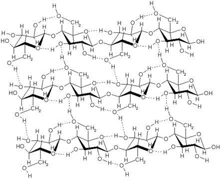
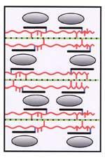
Primary cell made up of cellulose chains cross linked to each other; The primary cell is the first component to be deposited over the middle lamellae made up of pectic substances.
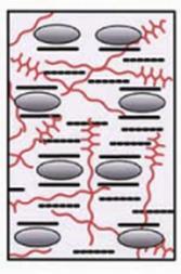
Glucoronoxylans, glucomannan, and feruloylated arabinoxylan form the hemicellulose components of secondary wall.


Plant cell are bound to each other by their cell walls, where the middle wall made up of pectate, which acts glue; this cell wall is traversed with many cytoplasmic strands called plasmodesmata.
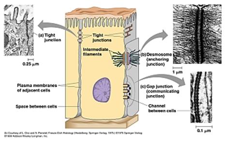
Animal cell are glued to each other with very many components such as tight junctions, gap junctions, desmosome elements. ; all of them are made up of cytoskeleton elements.

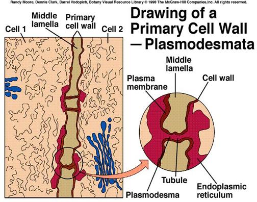
Plasmodesmata tubules interconnecting two cells.