FLUID TRANSPORT:
Loss of water I:
Guttation:
Inspite of water being an essential component of plant cells, instead of conserving, they lose water from their surfaces. By doing so they themselves get into peril, but they also benefit by this process. By the quirk of nature, plants have no control over the loss of water, because it is the environmental factors that force the plant to loose water in the form of water vapors or sometimes in liquid form. Nonetheless, many plants belonging to various taxons are adopted to check excessive loss of water and also conserve water. The process by which plants loose water in liquid form is called Guttation; on the other hand, if the water is lost from the plant body in the form of water vapors, then the phenomenon is called Transpiration, which is often referred to as an unavoidable evil process.
Guttation:
Plants which loose water by guttation are restricted to few taxons in the plant kingdom; hardly there are 150 – 250 plant species – ex. Tomato, grasses, colochasia, cucurbita members, balsam, etc. Colochasia, an aquatic plant has been found to loose more than 250 ml of water per day. Otherwise, the amount of water lost by guttation is hardly of any significance. Unlike transpiration process, guttation takes place only under certain conditions like high relative humidity in the atmosphere and plants with high amount of water in the soil.
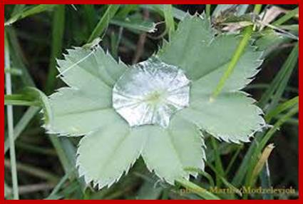
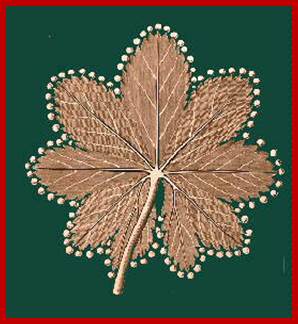
Alchemilla;-www.flowersinsweden.com; www.weezbo.com
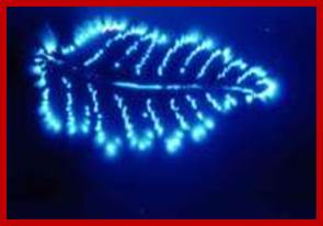
Naturally grown fresh fruits and vegetables, for example, are rich in biophotons. It’s obvious. You need not be a mystic who can see auras to understand. The reality of light waves, or biophotons energy, is obvious to any receptive and discerning eye.
Biophotons shown below, elevate the organism – such as your physical body – to a higher oscillation. As I read that, basically, if you eat fresh, clean food grown on healthy natural land, you support your body at a higher, healthier vibe.
https://thecalloftheland.wordpress.com
www.serenityinthegarden.blogspot.com; https://s10.lite.msu.edu

en.wikipedia.org; www.imagejuicy.com
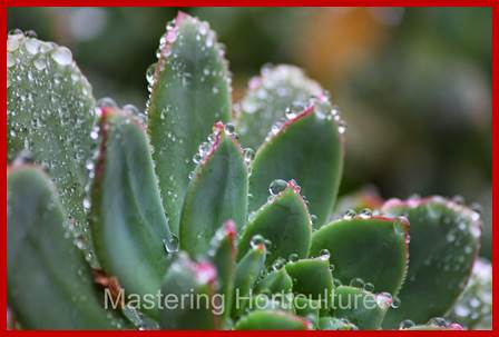
Water is pushed through Hydathodes; http://masteringhorticulture.blogspot.in/
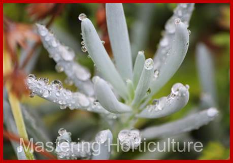
Senecio; Guttation in succulents; http://masteringhorticulture.blogspot.in/
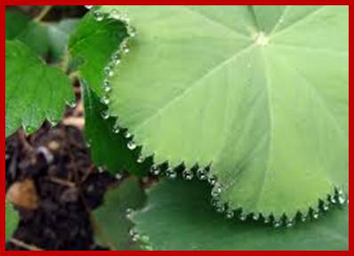
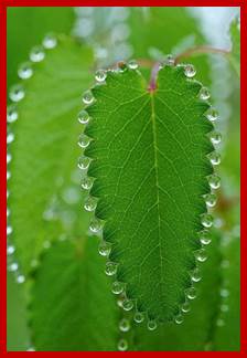
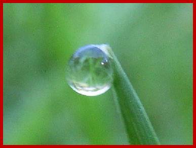
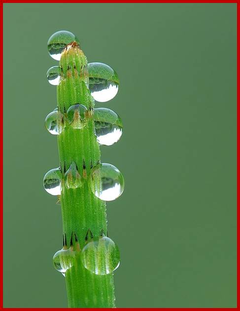
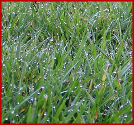
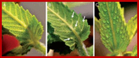
The water that is lost in this process is not pure but contains minerals, organic acids, sugars and even enzymes. The estimation of solutes lost in this process, reveal that certain plants loose about 200-500 mg of solutes per liter of water. On evaporation, the solutes that remain at the margins or tips of leaves cause salt burning, which is often called Guttation burn. The liquid drops that are exuded from the leaves are always through special structures called water stomata or hydathodes. Sometimes, the droplets found at the margin or apex, where hydathodes present, look like dew drops, but they are actually, the liquid drops that are forced out by guttation process.
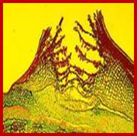
HYDATHODES
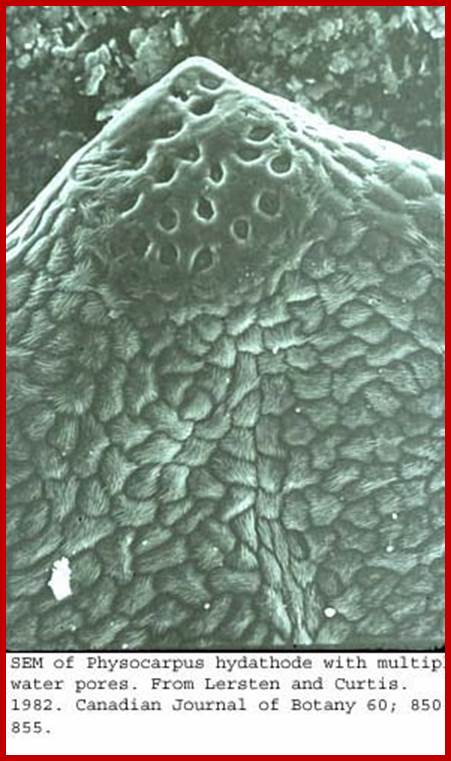
Hydathodes are specialized secretory tissue structures and they are mainly responsible for secreting water in liquid form. They are generally restricted to the apex or the serrated edges in the margins of leaves. They are mostly found in aquatic plants and in some herbaceous plants. These structures are connected to vascular bundles; it now believed they are evolved from modified stomata. This is also called ‘open water stomata or open Pore”
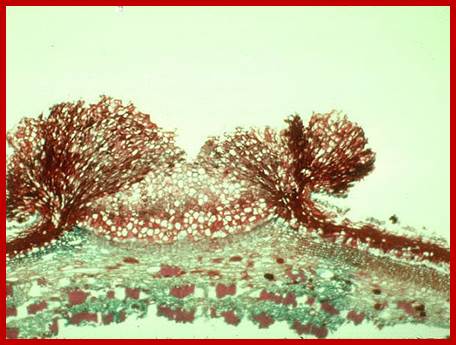
Longitudinal section of the leaf tip showing a mass of parenchymatous cells , at is base one finds vasculature terminal; primula sienensis; Brockhaus and Efron Encyclopedic Dictionary (Russian) http://www.biologydiscussion.com/
Rhinanthus alectorophus, a clade of Orobanchaceae possess metabolically active glandular trichomes that have been suggested to function as hydathode trichomes actively secreting water.
Root parasite; http://aob.oxfordjournals.org/
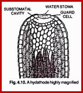
Structurally, hydathodes consist of a simple pore in the epidermal layer of cells found at the tip. In dorsi-ventral leaves they are found lower epidermal cells, but found on both surface in isobilateral leaves. A rhinanthoid clade of Orobanchaceae possess metabolically active glandular trichomes that have been suggested to function as hydathode-trichomes actively secreting water, a space found just behind the pore is surrounded by a special parenchymatous tissue. It is called Epithem. The cells are isodiametric in shape, loosely arranged and enclose a lot of intercellular spaces. An interesting feature is that the xylem elements of veinlets terminate in this tissue. Furthermore, some of the parenchymatous cells which surround the xylem elements exhibit structural features characteristic of transfer cells. Such cells contain a large number of finger shaped processes emanating from the cell wall, thus pushing the plasma membrane inwards into a similar pattern. These projections actually increase the surface area of the membranes considerably. Moreover, these cells are very active in transporting water and other cellular components. Thus, the epithem cells play a significant role in Guttation process.
Guttation can also cause injuries to plants-depletion of vital nutrients, burns at the tips of leaves and hydathodes can allow microorganisms that infect plants. The injury symptoms are chlorosis, necrosis due to action of concentrated solution that is supposed to be secreted. Epithem cells have the ability to retrieve nutrients from guttated liquid. Our previous studies (Chen and Chen, 2005; 2006) on the ultrastructure and morphogenesis of the laminar hydathodes of F. formosana showed that: 1, epithem cells have sinuous cell wall to increase their absorption surface area of cells; 2, both vigorous membrane morphological studies on laminar hydathodes of Ficus formosana (Moraceae) III. Salt injury of guttation on hydathodes Chyi-Chuann CHEN and Yung-Reui CHEN* Institute of Molecular and Cellular Biology, National Taiwan University, Taipei, Taiwan (Received October 20, 2005; Accepted September 8, 2006) ABSTRACT. The salt concentrations of gutted solution of laminar hydathodes on leaf usually increase after the repetition of guttation and eva-transpiration, and thus situation may lead to injure the hydathodes. The aim of this study is to investigate the salt injury of gutted solution on hydathodes of Ficus formosana Maxim.
Ultra structural studies show that the hypertonic stress of gutted solution caused by evaporation could lead the injury of hydathodes. The major symptoms of salt injury caused by hypertonic stress are as the follows: many electron-dense particles are spread in the nucleus and other organelles; the nucleolus is condensed and then disappeared; the endomembrane system is collapsed and then entirely become osmiophilic materials in the cytoplasm. Upon dehydration, the collapsed membranes become myelin-like structures are also observed. According to different degrees of salt injury within hydathodes, the abilities of tissue’s salt-tolerance are diversified and tolerance ability of the epithem is better than other tissues. These results imply that epithem possesses some special mechanisms that have been evolved to adapt the damage stress. In addition to physiological regulation, we suggest that some morphological changes such as the sinuous cell wall, proliferation of peroxisomes and the abundant endomembrane systems, and the conspicuous fluid-phase endocytosis.
Epithem promotes the tolerant efficiency of vacuoles by increasing the contact surface with environment to accelerate salt tolerance. Keywords: Epithem; Ficus formosana Maxim.; Fluid-phase endocytosis; Hydathodes; Sheath layer; Salt injury; Water pore. 216 Botanical Studies, Vol. 48, 2007 endocytosis and actively pumping endomembrane systems are induced by plasmolysis-de-plasmolysis cycles that support the membrane surface changes of epithem cells; 3, proliferated peroxisomes in epithem cells may depress the free radicals, which are produced by high salt stress; 4, observed many salt-glandular trichomes occur in the vicinity of hydathodes’ surface during the early stage of leaf development. Epithem cells couldn’t endure the strict stress circumstance coming even possess above characteristics, and the salt damage of membrane systems still happen (Kuchitsu et al., 1992; Hernandez et al., 1993; Huang, 1996). It is interesting to see whether these salt glandular trichomes have a function of removing and eliminating excess salt during leaf development. In this study, we tried to investigate the symptoms- http://ejournal.sinica.edu.tw/
Mechanism of Guttation:
Under certain conditions like soil flooded with overnight rain water and with high relative humidity of the day atmosphere, the root system of some plants like tomato, potato, etc., absorb excess of water by active uptake. As a result, hydrostatic pressure develops in the root system which actually pushes water upwards. So the water along with other soluble components of the cells is forced out of the xylem elements located into Epithem tissue. As result, the space behind the water stomata gets filled with the water and with more root pressure operating; the liquid is virtually pushed out of the pore, where the stomata do not offer any resistance. Probably transfer cells may also help in the retrieval of minerals and other components from the xylem elements and secreting out along with water. However, it has been speculated that active hydathodes may directly secrete the minerals and organic acids out of the passive stomata. Such active secretion of the above said substances creates a diffusion gradient and water is just withdrawn from the cells into exterior surface so guttation takes place. In spite of the borderline between active and passive mechanisms of guttation is not much, the concepts are attractive.
www.plantphys.info
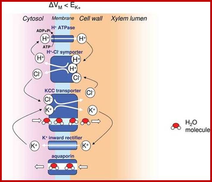
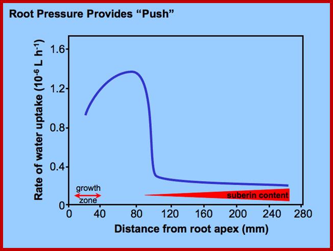
The thermodynamics of root pressure remains an enigma up to the present day. Water is transported radially into xylem vessels, under some conditions even when the xylem sap is more dilute than the ambient medium (soil solution). It is suggested here that water secretion across the plasma membrane of xylem parenchyma cells is driven by a co-transport of water and solutes as previously shown for mammalian epithelia (Zeuthen T. 2010. Water-transporting proteins. Journal of Membrane Biology 234, 57–73.). This process could drive volume flow ‘energetically uphill’, against the free energy gradient of water. According to the model, solutes released by xylem parenchyma cells are subsequently retrieved from the sap at the expense of metabolic energy to maintain the concentration gradient that drives the water secretion. Transporters of the CCC type known to mediate water secretion in mammalian cells have also been found in Arabidopsis and in rice. The mechanism proposed here for root pressure could also explain refilling of embolized vessels. Moreover, it could contribute to long-distance water transport in trees when the cohesion–tension mechanism of water ascent fails. This is discussed with respect to the old and the more recent literature on these subjects. Key words: Aquaporin, CCC transporters, cohesion–tension (CT) theory, embolism repair, reflection coefficient, root pressure, water ascent, water co-transport hypothesis, xylem refilling. Journal of Experimental Botany, Vol. 65, No. 2, pp. 381–393, 2014 doi:10.1093/jxb/ert391 Advance Access publication 5 December, 2013 Opinion Paper Root pressure and beyond: energetically uphill water transport into xylem vessels? Journal of Experimental Botany, Vol. 65, No. 2, pp. 381–393, 2014.
Root pressure causes hydathodes to ooze out water. When soil is flooded with overnight rain and high relative humidity roots of like tomato plant and others absorb excess water by active uptake and develop hydrostatic pressure or call it as root pressure that pushes the upwards or say forced out. So the water with soluble components is pushed out hydathodes.
Lenticular Transpiration:
Lenticels are considered as the breathing pores of the bark. Whichever plants that exhibit secondary growth, produce lenticels in stems as well as in roots. During periderm formation, the cork cambium at certain regions, instead of producing phellum starts producing a group of parenchymatous cells called complementary cells. They are isodiametric, rich in cytoplasm, loosely arranged and thin walled. As the complementary tissue is locally produced, the epidermis is pushed out and finally it breaks open thus exposes the complementary tissue to the external atmosphere.
As the complementary tissue contains so much of intercellular spaces, the atmospheric air diffuses easily into them. Depending upon the relative humidity of atmospheric air, water from complementary cells is lost to intercellular spaces, from which it finds its way into the atmosphere. This process takes place throughout the day and night and there is no way by which it can be stopped. Fortunately, the total amount of water lost by this method is again not that significant. It is not uncommon to see, the loss of water through the bark. This happens in spite of suberization of bark cells. However, bark transpiration, along with lenticular transpiration do not cause any serious injury to plants.
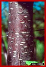
Lenticels or breathing pores; www.cas.miamioh.edu
Sucrose transports SUT 1; 12 predicted transmembrane domains, and are assumed to form a single pore for sucrose, with N- and C-termini and 5 even-numbered loops located in the cytosol.